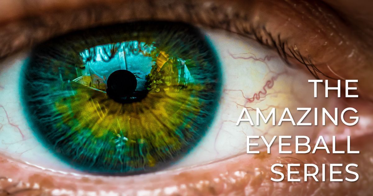
The Amazing Eyeball: Part 8 - The Optic Nerve

October 9, 2024
A close up of a person 's eye with the words

Welcome to The Amazing Eyeball, a comprehensive 10-part series exploring the intricate structures that make up one of the body’s most remarkable organs - the human eye. Each article in this series delves deep into the anatomy and function of different parts of the eye. Throughout the series, we’ll uncover how these structures work together to produce the miracle of sight, along with insights into common eye conditions, cutting-edge treatments, and the eye’s natural healing abilities. Whether you're fascinated by the eye's biology or eager to learn how to protect your vision, this series will take you on a journey through the wonders of the human eye.


The Optic Nerve: Your Eye’s Direct Line to the Brain

Read time: 7 minutes


While the eye captures light and processes visual information, it’s the optic nerve that carries these signals to the brain, where they are transformed into the images we see. The optic nerve serves as the vital connection between the eye and the brain, making it essential for vision. Without this direct line of communication, the eye would be unable to convey the detailed information it gathers to the brain, leaving us blind to the world around us.


In this article, we will explore the anatomy and function of the optic nerve, its role in vision, and the conditions that can affect its health. This piece is part of our ongoing series on the makeup of the eyeball. Be sure to check out our previous articles on the retina, cornea, aqueous and vitreous humor, iris and pupil, lens, macula and fovea, and sclera to understand how the optic nerve fits into the complex system that allows us to see.


What is the Optic Nerve?


The optic nerve is a bundle of over 1 million nerve fibers that transmit visual information from the retina to the brain. It is considered part of the central nervous system (CNS) rather than the peripheral nervous system, due to its direct connection with the brain.


The optic nerve is composed primarily of the axons of the retinal ganglion cells, which collect visual signals from the retina. These signals, which begin as light hitting the retina, are converted into electrical impulses by photoreceptor cells and then transmitted through the optic nerve to the brain’s visual cortex, where they are processed into meaningful images.


Key features of the optic nerve include:


  • Length: The optic nerve spans approximately 4 cm (1.6 inches) from the back of the eye to the brain.


  • Optic Disc: The optic nerve begins at the optic disc, a small circular area at the back of the eye where the nerve fibers converge to form the optic nerve. The optic disc is also known as the blind spot, as there are no photoreceptor cells in this region to detect light.


  • Crossing at the Optic Chiasm: The optic nerve from each eye meets at a structure called the optic chiasm, where some of the nerve fibers cross over to the opposite side of the brain. This crossing allows visual information from both eyes to be integrated and processed together, contributing to depth perception and a full field of vision.


How the Optic Nerve Transmits Visual Information


The optic nerve is essentially the visual information highway that carries signals from the retina to the brain. Here’s how this process works:


  1. Light enters the eye and is focused onto the retina, where photoreceptor cells (rods and cones) detect the light and convert it into electrical signals.
  2. These electrical signals are transmitted to the retinal ganglion cells, whose axons make up the optic nerve.
  3. The optic nerve carries the signals from the retina to the optic chiasm, where some fibers cross over to the opposite hemisphere of the brain.
  4. After crossing the optic chiasm, the signals are relayed through the optic tracts to the lateral geniculate nucleus (LGN) in the brain, and from there, they are sent to the visual cortex in the occipital lobe.
  5. The brain processes these signals, transforming them into the images we perceive, allowing us to understand the visual information in our surroundings.


This entire process happens in fractions of a second, allowing us to see the world in real-time.


The Optic Disc and the Blind Spot


As mentioned earlier, the optic disc is the point where the optic nerve begins. This is also known as the blind spot because it lacks photoreceptor cells, meaning it cannot detect light. Normally, the brain compensates for this small gap in vision by filling in the missing information based on surrounding details and input from the other eye.


The optic disc is also the entry and exit point for blood vessels that supply the retina, making it an essential structure for both visual transmission and the eye’s nourishment.


Conditions Affecting the Optic Nerve


Because the optic nerve is essential for transmitting visual information, damage to the optic nerve can lead to significant vision problems, including blindness. Several conditions can affect the optic nerve, either through direct injury or as a result of systemic health issues.


Glaucoma


One of the most common and serious conditions affecting the optic nerve is glaucoma. Glaucoma occurs when increased pressure inside the eye, known as intraocular pressure (IOP), damages the optic nerve over time. As the optic nerve fibers are progressively damaged, blind spots develop in the peripheral vision, and if untreated, glaucoma can lead to complete vision loss.


There are two primary types of glaucoma:


  • Open-angle glaucoma: The most common form, where fluid drains too slowly from the eye, increasing pressure gradually.


  • Angle-closure glaucoma: A less common but more severe form, where the drainage angle between the iris and cornea becomes blocked, causing a rapid increase in pressure.


Early detection through regular eye exams is crucial for managing glaucoma, as the damage it causes to the optic nerve is irreversible. Treatment often involves medications or surgery to reduce intraocular pressure and slow the progression of vision loss.


Optic Neuritis


Optic neuritis is an inflammation of the optic nerve that can cause sudden vision loss, typically in one eye. This condition is often associated with multiple sclerosis (MS), an autoimmune disease that attacks the central nervous system, including the optic nerve.


Symptoms of optic neuritis include:


  • Blurred or dimmed vision.
  • Pain in or around the eye, especially during eye movement.
  • Loss of color vision or a reduction in contrast.


Optic neuritis often improves on its own, but corticosteroids may be used to reduce inflammation and speed up recovery. However, if the underlying cause is MS or another autoimmune condition, further treatment may be required to manage the disease.


Optic Neuropathy


Optic neuropathy refers to damage to the optic nerve, often resulting in vision loss. This damage can be caused by various factors, including reduced blood flow (known as ischemic optic neuropathy), trauma, or toxic substances.


In some cases, optic neuropathy can be the result of underlying conditions like diabetes, hypertension, or certain medications that damage the optic nerve over time.


Optic Atrophy


Optic atrophy is the degeneration of the optic nerve fibers, leading to vision loss. This condition can result from a variety of causes, including glaucoma, optic neuritis, or trauma to the optic nerve. Optic atrophy is characterized by the loss of visual acuity and contrast sensitivity, and it can affect both central and peripheral vision.

Unfortunately, optic atrophy is usually irreversible, as the nerve fibers cannot regenerate once they are damaged. Treatment typically focuses on managing the underlying cause to prevent further vision loss.


Papilledema


Papilledema is a condition where the optic disc swells due to increased pressure within the skull, often caused by conditions such as brain tumors, head trauma, or severe hypertension. This swelling can compress the optic nerve and lead to vision problems such as blurred vision, double vision, or temporary vision loss.

Papilledema requires prompt medical attention, as it can indicate a serious underlying condition affecting the brain. Treating the root cause of the increased intracranial pressure is essential to prevent permanent damage to the optic nerve.


How to Protect the Optic Nerve


Maintaining the health of your optic nerve is crucial for preserving your vision. Here are some steps you can take to protect this vital structure:


  1. Regular Eye Exams: Comprehensive eye exams can detect early signs of optic nerve damage, such as glaucoma or optic neuropathy, before significant vision loss occurs. If you are at higher risk for optic nerve conditions (due to family history or other health conditions), regular checkups are especially important.
  2. Manage Chronic Conditions: Diseases such as diabetes, hypertension, and autoimmune disorders can damage the optic nerve if not properly managed. Work with your healthcare provider to control these conditions and reduce the risk of complications affecting your eyes.
  3. Avoid Smoking: Smoking has been linked to an increased risk of optic neuropathy, as it can restrict blood flow to the optic nerve. Quitting smoking can significantly improve your overall eye health.
  4. Protect Your Head: Head injuries can lead to damage to the optic nerve, particularly if the injury causes swelling or trauma around the eye or brain. Always wear appropriate protective gear during sports or high-risk activities to reduce the risk of head trauma.
  5. Control Eye Pressure: If you have glaucoma or are at risk for developing it, follow your doctor’s treatment plan to control intraocular pressure and prevent damage to the optic nerve.


The Takeaway


The optic nerve is a critical part of the visual system, responsible for carrying the signals generated by the retina to the brain, where they are processed into the images we see. Its health is vital for vision, and damage to the optic nerve can lead to significant vision loss. By taking steps to protect your optic nerve and staying vigilant about regular eye care, you can help preserve your vision for years to come.


In the next article in our series, we will explore the choroid, the layer of blood vessels that nourishes the retina and other parts of the eye. Stay tuned as we continue our journey through the anatomy of the eyeball!


Read the next article in this series: The Amazing Eyeball: Part 9 - The Choroid

Share this blog post on social or with a friend:

The information provided in this article is intended for general knowledge and educational purposes only and should not be construed as medical advice. It is strongly recommended to consult with an eye care professional for personalized recommendations and guidance regarding your individual needs and eye health concerns.


All of Urban Optiks Optometry's blog posts and articles contain information carefully curated from openly sourced materials available in the public domain. We strive to ensure the accuracy and relevance of the information provided. For a comprehensive understanding of our practices and to read our full disclosure statement, please click here.

OUR LATEST POSTS

Woman wearing yellow-rimmed sunglasses and a cream-colored top with arms above her head.
October 22, 2025
Discover why some designs endure in fashion. Explore how eyewear luxury brands like Urban Optiks Optometry blend style with timeless quality.
Woman in profile view looking off in the distance, with a blurred person in the background.
October 17, 2025
Discover how eye movement patterns reveal emotional intelligence. Learn the science behind gaze, cultural variations, and how eye-tracking research enhances communication, leadership, and emotional skills.
Man in plaid blazer and sunglasses, standing outdoors with autumn foliage.
October 12, 2025
Discover handcrafted eyewear designed for San Diego’s fall lifestyle. Explore artisan frames at Urban Optiks Optometry that blend comfort and style.
Close-up of an eye with an abrasion, illuminated with ultraviolet light.
October 7, 2025
Read time: 5 minutes The cornea is the clear, dome-shaped surface at the front of the eye. It plays two essential roles: focusing light for sharp vision and protecting the eye’s delicate inner structures from debris and infection. Because it is both exposed and highly sensitive, the cornea is especially vulnerable to injury. A scratch or scrape on its surface is known as a corneal abrasion. While many abrasions are minor and heal quickly, they can cause significant discomfort and may lead to complications if left untreated. Knowing the causes, symptoms, and treatments can help you protect your vision and respond appropriately if an injury occurs. What Is a Corneal Abrasion? A corneal abrasion is a scratch or scrape on the epithelium, the thin outer layer of the cornea. This surface contains numerous nerve endings, which explains why even small abrasions often cause sudden, sharp pain. Along with discomfort, abrasions can also blur vision until the surface has healed. You can find out more about corneal abrasions in this article from Harvard Health Publishing . Common Causes Corneal abrasions often happen in everyday life, from something as simple as rubbing the eye too hard to environmental hazards at work or outdoors. Some of the most common causes include: Foreign particles such as dust, sand, or metal shavings entering the eye Accidental scratches , often from fingernails or makeup brushes Improper contact lens use , including wearing them overnight or with poor hygiene Paper cuts from envelopes or books Tree branches or leaves encountered outdoors Workplace hazards , especially when protective eyewear is not used Symptoms to Watch For Because the cornea is so sensitive, symptoms usually appear immediately. These may include: Sudden eye pain, especially noticeable when blinking Redness, tearing, and excessive blinking A persistent sensation of something in the eye (foreign body sensation) Light sensitivity ( photophobia ) Blurred or decreased vision Difficulty keeping the eye open These symptoms can overlap with other conditions, including corneal infections, so a professional exam is recommended rather than self-diagnosis. Diagnosis Eye doctors typically use a slit-lamp microscope to examine the cornea in detail. They may also apply a fluorescein dye, which makes scratches more visible under blue light. This exam helps confirm the abrasion and check for any trapped foreign objects. In more serious cases, or if caused by high-velocity particles, additional imaging may be used. Treatment and Healing Most corneal abrasions heal within 24 to 72 hours. Treatment focuses on pain relief, preventing infection, and supporting the natural healing process. Depending on the injury, your doctor may recommend: Lubricating drops or ointments to ease irritation Antibiotic drops to prevent infection (especially important for contact lens wearers) Prescription drops to relax eye muscles and reduce pain in some cases Avoiding contact lenses until healing is complete Protective eyewear to prevent further trauma Eye patches are rarely used today, and steroid drops are generally avoided during the initial healing period since they can slow recovery. Special Considerations for Contact Lens Wearers Contact lenses increase the risk of both abrasions and infections. Wearing lenses too long, handling them without proper hygiene, or sleeping in them can scratch the cornea and create conditions for bacterial growth. If you wear contacts and develop sudden eye pain, redness, or blurry vision, remove the lenses immediately and see your eye doctor. Continuing to wear contacts over an abrasion can worsen the injury and delay healing. When to Seek Medical Care Even if symptoms seem mild, it’s best to have corneal abrasions evaluated by an optometrist or ophthalmologist. Seek prompt care if: Pain lasts longer than 24 hours Vision becomes increasingly blurry Sensitivity to light worsens There is discharge, swelling, or signs of infection You have a history of recurrent corneal erosions Without treatment, abrasions may lead to complications such as corneal ulcers or scarring, both of which can cause permanent vision changes. Prevention  Many abrasions are preventable with good habits. Steps you can take include: Wearing protective eyewear during high-risk activities like construction, sports, or yard work Handling contact lenses with clean hands and avoiding overnight wear unless prescribed Replacing eye makeup tools regularly and keeping them clean Avoiding rubbing your eyes, especially in dusty or dry environments Using artificial tears to keep the eyes hydrated The Takeaway Corneal abrasions may be common, but they should never be ignored. With proper treatment, most heal quickly and without lasting effects. However, untreated abrasions can progress into serious eye conditions that permanently affect vision. Whether from contact lenses, workplace exposure, or everyday accidents, protecting your cornea is essential to safeguarding your eyesight. Learn more about other corneal conditions in this article from the National Eye Institute.
Woman with dark hair squinting at sunglasses with a concerned expression.
October 2, 2025
Learn what causes eyewear allergies, common frame material triggers, and hypoallergenic alternatives
Doctor holding a brain model, showing a patient, other doctors looking at scan in background.
September 27, 2025
Learn how strokes can affect vision, from field loss to eye movement issues, and discover strategies that support adaptation and visual independence.
Woman in sunglasses with shopping bags over her shoulders is smiling while outdoors.
September 22, 2025
Explore how touch, sight, and feel impact your fashion choices. Visit Urban Optiks Optometry in San Diego for an immersive sensory shopping experience.
Two women in sunglasses and fashionable accessories, smiling and posing together.
September 15, 2025
Discover how accessories and confidence transform style. Explore luxury sunglasses brands to elevate your look with Urban Optiks Optometry.
Colorfully dressed young adults posing as a photographer takes their picture.
September 11, 2025
Explore how pop culture influences your style. Discover practical tips, including contact lenses in San Diego, to enhance your everyday fashion.
A collage of images of people wearing glasses
September 4, 2025
Uncover how your personal style choices in luxury designer eyewear reflect your personality. Explore the latest trends at Urban Optiks Optometry.
Woman wearing yellow-rimmed sunglasses and a cream-colored top with arms above her head.
October 22, 2025
Discover why some designs endure in fashion. Explore how eyewear luxury brands like Urban Optiks Optometry blend style with timeless quality.
Woman in profile view looking off in the distance, with a blurred person in the background.
October 17, 2025
Discover how eye movement patterns reveal emotional intelligence. Learn the science behind gaze, cultural variations, and how eye-tracking research enhances communication, leadership, and emotional skills.
Man in plaid blazer and sunglasses, standing outdoors with autumn foliage.
October 12, 2025
Discover handcrafted eyewear designed for San Diego’s fall lifestyle. Explore artisan frames at Urban Optiks Optometry that blend comfort and style.
Close-up of an eye with an abrasion, illuminated with ultraviolet light.
October 7, 2025
Read time: 5 minutes The cornea is the clear, dome-shaped surface at the front of the eye. It plays two essential roles: focusing light for sharp vision and protecting the eye’s delicate inner structures from debris and infection. Because it is both exposed and highly sensitive, the cornea is especially vulnerable to injury. A scratch or scrape on its surface is known as a corneal abrasion. While many abrasions are minor and heal quickly, they can cause significant discomfort and may lead to complications if left untreated. Knowing the causes, symptoms, and treatments can help you protect your vision and respond appropriately if an injury occurs. What Is a Corneal Abrasion? A corneal abrasion is a scratch or scrape on the epithelium, the thin outer layer of the cornea. This surface contains numerous nerve endings, which explains why even small abrasions often cause sudden, sharp pain. Along with discomfort, abrasions can also blur vision until the surface has healed. You can find out more about corneal abrasions in this article from Harvard Health Publishing . Common Causes Corneal abrasions often happen in everyday life, from something as simple as rubbing the eye too hard to environmental hazards at work or outdoors. Some of the most common causes include: Foreign particles such as dust, sand, or metal shavings entering the eye Accidental scratches , often from fingernails or makeup brushes Improper contact lens use , including wearing them overnight or with poor hygiene Paper cuts from envelopes or books Tree branches or leaves encountered outdoors Workplace hazards , especially when protective eyewear is not used Symptoms to Watch For Because the cornea is so sensitive, symptoms usually appear immediately. These may include: Sudden eye pain, especially noticeable when blinking Redness, tearing, and excessive blinking A persistent sensation of something in the eye (foreign body sensation) Light sensitivity ( photophobia ) Blurred or decreased vision Difficulty keeping the eye open These symptoms can overlap with other conditions, including corneal infections, so a professional exam is recommended rather than self-diagnosis. Diagnosis Eye doctors typically use a slit-lamp microscope to examine the cornea in detail. They may also apply a fluorescein dye, which makes scratches more visible under blue light. This exam helps confirm the abrasion and check for any trapped foreign objects. In more serious cases, or if caused by high-velocity particles, additional imaging may be used. Treatment and Healing Most corneal abrasions heal within 24 to 72 hours. Treatment focuses on pain relief, preventing infection, and supporting the natural healing process. Depending on the injury, your doctor may recommend: Lubricating drops or ointments to ease irritation Antibiotic drops to prevent infection (especially important for contact lens wearers) Prescription drops to relax eye muscles and reduce pain in some cases Avoiding contact lenses until healing is complete Protective eyewear to prevent further trauma Eye patches are rarely used today, and steroid drops are generally avoided during the initial healing period since they can slow recovery. Special Considerations for Contact Lens Wearers Contact lenses increase the risk of both abrasions and infections. Wearing lenses too long, handling them without proper hygiene, or sleeping in them can scratch the cornea and create conditions for bacterial growth. If you wear contacts and develop sudden eye pain, redness, or blurry vision, remove the lenses immediately and see your eye doctor. Continuing to wear contacts over an abrasion can worsen the injury and delay healing. When to Seek Medical Care Even if symptoms seem mild, it’s best to have corneal abrasions evaluated by an optometrist or ophthalmologist. Seek prompt care if: Pain lasts longer than 24 hours Vision becomes increasingly blurry Sensitivity to light worsens There is discharge, swelling, or signs of infection You have a history of recurrent corneal erosions Without treatment, abrasions may lead to complications such as corneal ulcers or scarring, both of which can cause permanent vision changes. Prevention  Many abrasions are preventable with good habits. Steps you can take include: Wearing protective eyewear during high-risk activities like construction, sports, or yard work Handling contact lenses with clean hands and avoiding overnight wear unless prescribed Replacing eye makeup tools regularly and keeping them clean Avoiding rubbing your eyes, especially in dusty or dry environments Using artificial tears to keep the eyes hydrated The Takeaway Corneal abrasions may be common, but they should never be ignored. With proper treatment, most heal quickly and without lasting effects. However, untreated abrasions can progress into serious eye conditions that permanently affect vision. Whether from contact lenses, workplace exposure, or everyday accidents, protecting your cornea is essential to safeguarding your eyesight. Learn more about other corneal conditions in this article from the National Eye Institute.
Woman with dark hair squinting at sunglasses with a concerned expression.
October 2, 2025
Learn what causes eyewear allergies, common frame material triggers, and hypoallergenic alternatives