The Amazing Eyeball: Part 7 - The Sclera

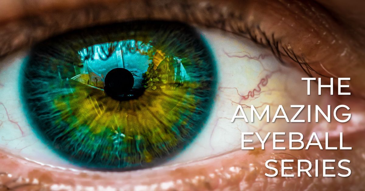
Welcome to The Amazing Eyeball, a comprehensive 10-part series exploring the intricate structures that make up one of the body’s most remarkable organs - the human eye. Each article in this series delves deep into the anatomy and function of different parts of the eye. Throughout the series, we’ll uncover how these structures work together to produce the miracle of sight, along with insights into common eye conditions, cutting-edge treatments, and the eye’s natural healing abilities. Whether you're fascinated by the eye's biology or eager to learn how to protect your vision, this series will take you on a journey through the wonders of the human eye.
The Sclera: The White of the Eye and Its Protective Function
Read time: 7 minutes
When we think about the anatomy of the eye, the focus often lies on the more noticeable components like the cornea, retina, and lens. However, one of the most important parts of the eye, providing structural support and protection, is the sclera—the white outer layer that we commonly associate with the visible “white of the eye.” The sclera plays a vital role in maintaining the eye’s shape and shielding its delicate internal structures from injury.
In this article, we will explore the structure and function of the sclera, its importance for eye health, and the conditions that can affect it. This piece is part of our ongoing series on the makeup of the eyeball. Be sure to check out our previous articles on the retina, cornea, aqueous and vitreous humor, iris and pupil, lens, and macula and fovea to get a complete understanding of how these components work together to support healthy vision.
The Structure of the Sclera
The sclera is the tough, fibrous outer layer of the eye that forms about 80% of the eyeball’s surface area. It is composed of dense connective tissue and serves several essential functions, including providing structural support and protecting the inner parts of the eye from injury and environmental factors.
The sclera has a distinct white color in most people, although in newborns and some individuals with specific medical conditions, it may appear blue-tinted or slightly yellowish.
Key structural components of the sclera include:
- Collagen and Elastic Fibers: The sclera’s tough composition comes from its high concentration of collagen fibers, which give it strength and maintain the shape of the eye. Elastic fibers allow for flexibility, enabling the eye to move without losing its structural integrity.
- Thickness: The sclera varies in thickness throughout different parts of the eye. It is thickest at the back, near the optic nerve, and thinnest at the front, where it connects to the cornea.
The Sclera’s Protective Role
The primary function of the sclera is to protect the eye from external damage. Its tough, durable nature acts as a shield, preventing injury to the sensitive inner structures, including the retina, lens, and optic nerve. The sclera also helps maintain the shape of the eyeball, ensuring that light is properly focused on the retina for clear vision.
Additionally, the sclera provides a point of attachment for the extraocular muscles, which are responsible for moving the eye in different directions. These muscles are anchored to the sclera, allowing the eye to rotate smoothly and adjust its gaze.
Scleral Blood Supply and Nourishment
Although the sclera appears avascular (lacking blood vessels), it actually receives nourishment from blood vessels in the surrounding tissues, particularly from the episclera and the choroid. The episclera is a thin, vascular layer that lies on top of the sclera and supplies it with oxygen and nutrients through small blood vessels.
The choroid, which lies beneath the sclera, also plays a role in nourishing the eye. This vascular layer is rich in blood vessels and provides the retina and other inner structures with the oxygen and nutrients they need to function properly.
Sclera vs. Cornea: A Clear Distinction
Although the sclera and cornea are both outer layers of the eye, they serve very different purposes. While the sclera is opaque and provides structural support and protection, the cornea is transparent and responsible for allowing light to enter the eye.
The transition between the sclera and cornea occurs at a region known as the limbus, where the white of the eye meets the clear, dome-shaped cornea. While the sclera consists mainly of collagen, the cornea’s structure is more specialized to maintain clarity and transparency, allowing light to pass through to the lens and retina.
Common Conditions Affecting the Sclera
While the sclera is tough and resilient, it can be affected by several conditions that may compromise its function or appearance. Some of the most common conditions include:
Scleritis
Scleritis is an inflammation of the sclera that can cause redness, pain, and vision problems. This condition is often associated with autoimmune diseases such as rheumatoid arthritis or lupus, where the body’s immune system mistakenly attacks healthy tissues.
Symptoms of scleritis include:
- Severe eye pain that may worsen with movement.
- Redness of the eye, particularly deep within the sclera.
- Blurred or reduced vision.
- Sensitivity to light (photophobia).
Scleritis requires prompt medical attention, as untreated inflammation can lead to complications such as thinning of the sclera, retinal detachment, or even vision loss. Treatment typically involves anti-inflammatory medications or corticosteroids to reduce inflammation and address the underlying autoimmune condition.
Episcleritis
Episcleritis is a less severe inflammation of the episclera, the layer of tissue that covers the sclera. Unlike scleritis, episcleritis is usually mild and self-limiting, often resolving on its own without treatment.
Symptoms of episcleritis include:
- Redness or swelling of the eye.
- Mild discomfort or tenderness.
- No significant vision changes.
Episcleritis may be associated with underlying conditions like allergies, infections, or autoimmune disorders, but it is generally a benign condition. If needed, treatment may involve lubricating eye drops or anti-inflammatory medications to alleviate symptoms.
Blue Sclera
Blue sclera is a condition where the sclera appears bluish due to its thinness. This thinning allows the underlying choroid to show through, giving the sclera a blue tint. Blue sclera can occur as a result of various genetic disorders, such as osteogenesis imperfecta, which affects the body’s ability to produce collagen.
In some cases, blue sclera may be a normal variation, particularly in infants, but it can also indicate an underlying connective tissue disorder that requires medical evaluation.
Scleral Ectasia
Scleral ectasia is a condition in which the sclera thins and bulges outward, often due to trauma, surgery, or disease. This thinning weakens the structural integrity of the eye and can lead to complications such as uveal prolapse (where the internal tissues of the eye bulge through the thinned sclera).
In severe cases, scleral ectasia may require surgical intervention to reinforce the weakened area and prevent further damage.
Surgical and Cosmetic Applications Involving the Sclera
In addition to its protective role, the sclera is sometimes involved in surgical or cosmetic procedures:
- Scleral Buckling: Scleral buckling is a surgical procedure used to treat retinal detachment. In this procedure, a flexible band is placed around the outside of the eye (on the sclera) to gently push the wall of the eye inward, allowing the detached retina to reattach to the underlying tissue. This procedure is often highly successful in preventing permanent vision loss caused by retinal detachment.
- Cosmetic Eye Whitening (Scleral Tattooing): Some cosmetic procedures involve altering the appearance of the sclera. For instance, scleral tattooing is a controversial practice in which ink is injected into the sclera to change its color. This procedure is not medically necessary and can carry significant risks, including infection, inflammation, and vision loss. Additionally, cosmetic eye whitening procedures aim to remove discoloration or blood vessels from the sclera to create a whiter, brighter appearance. While these procedures are often sought for aesthetic reasons, they should be approached with caution, as they can carry risks to eye health.
How to Protect the Sclera
Maintaining the health of the sclera is essential for overall eye health and vision. Here are some steps you can take to protect your sclera:
- Wear Protective Eyewear: Whether working in hazardous environments, participating in sports, or spending time outdoors, wearing protective eyewear can shield your eyes from injury and exposure to harmful elements like dust, chemicals, and UV rays.
- Avoid Rubbing Your Eyes: Rubbing your eyes can irritate the sclera and lead to conditions like episcleritis or worsen existing inflammation. If your eyes are itchy or uncomfortable, try using lubricating eye drops or antihistamines.
- Manage Underlying Health Conditions: If you have an autoimmune disorder or other chronic health condition, work with your healthcare provider to manage your symptoms and reduce the risk of complications that could affect your eyes.
- Regular Eye Exams: Routine eye exams can detect early signs of scleral conditions, such as thinning or inflammation. Early intervention can help prevent complications and preserve your vision.
The Takeaway
The sclera may not get as much attention as other parts of the eye, but it plays a crucial role in protecting and supporting the delicate structures that allow us to see. By maintaining the health of your sclera and seeking treatment for any inflammatory conditions, you can help ensure the overall well-being of your eyes.
In the next article in our series, we’ll explore the optic nerve, the eye’s direct connection to the brain, and how it transmits visual information. Stay tuned as we continue our journey through the anatomy of the eyeball!
Read the next article in this series: The Amazing Eyeball: Part 8 - The Optic Nerve
Share this blog post on social or with a friend:
The information provided in this article is intended for general knowledge and educational purposes only and should not be construed as medical advice. It is strongly recommended to consult with an eye care professional for personalized recommendations and guidance regarding your individual needs and eye health concerns.
All of Urban Optiks Optometry's blog posts and articles contain information carefully curated from openly sourced materials available in the public domain. We strive to ensure the accuracy and relevance of the information provided. For a comprehensive understanding of our practices and to read our full disclosure statement, please click here.


