The Amazing Eyeball: Part 9 - The Choroid
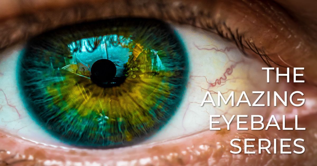
Welcome to The Amazing Eyeball, a comprehensive 10-part series exploring the intricate structures that make up one of the body’s most remarkable organs - the human eye. Each article in this series delves deep into the anatomy and function of different parts of the eye. Throughout the series, we’ll uncover how these structures work together to produce the miracle of sight, along with insights into common eye conditions, cutting-edge treatments, and the eye’s natural healing abilities. Whether you're fascinated by the eye's biology or eager to learn how to protect your vision, this series will take you on a journey through the wonders of the human eye.
The Choroid: The Eye’s Vascular Network for Nourishment and Health
Read time: 7 minutes
While many people are familiar with the retina, cornea, and lens, fewer are aware of the critical role the choroid plays in maintaining eye health. The choroid is a layer of blood vessels and connective tissue located between the sclera (the white of the eye) and the retina. Its primary function is to provide oxygen and nutrients to the retina, the light-sensitive tissue at the back of the eye that processes visual information.
In this article, we’ll explore the anatomy and function of the choroid, its role in nourishing the eye, and the conditions that can affect its health. This piece is part of our ongoing series on the makeup of the eyeball. Be sure to explore our previous articles on the retina, cornea, aqueous and vitreous humor, iris and pupil, lens, macula and fovea, sclera, and optic nerve to get a complete understanding of how these structures work together for optimal vision.
The Anatomy of the Choroid
The choroid is a thin, vascular layer sandwiched between the sclera and retina. It is primarily composed of blood vessels, providing essential nutrients to the outer layers of the retina, particularly the retinal pigment epithelium (RPE) and the photoreceptor cells.
The choroid is densely packed with melanocytes, which are cells that produce the pigment melanin. This pigment helps reduce light scatter within the eye, improving the clarity and sharpness of vision. The rich blood supply in the choroid allows it to nourish the retina and support its complex functions.
The choroid is divided into several layers:
- Haller’s Layer: The outermost layer, which contains large blood vessels.
- Sattler’s Layer: The middle layer, composed of medium-sized blood vessels.
- Choriocapillaris: The innermost layer, which is a network of fine capillaries that supplies oxygen and nutrients to the retina and retinal pigment epithelium (RPE).
The Function of the Choroid: Nourishment and Protection
The choroid’s primary role is to supply the outer part of the retina with oxygen and nutrients while removing waste products. Since the retina is one of the most metabolically active tissues in the body, it requires a constant and rich supply of oxygen to function properly.
Key functions of the choroid include:
- Oxygen and Nutrient Supply: The retina relies on the choroid to supply essential oxygen and nutrients. Without this vascular network, the photoreceptor cells in the retina would not have the resources necessary to convert light into electrical signals.
- Waste Removal: The choroid also plays a critical role in removing waste products from the retina, ensuring the retinal cells remain healthy and functional.
- Absorption of Light: The pigment in the choroid absorbs excess light that enters the eye, reducing light scatter and preventing glare. This helps improve the sharpness of the image projected onto the retina.
How the Choroid Supports the Retina
The health of the retina, and by extension our ability to see, depends heavily on the choroid’s ability to maintain a stable supply of oxygen and nutrients. The choriocapillaris, the thin layer of capillaries within the choroid, is responsible for supplying blood to the outer part of the retina, particularly the retinal pigment epithelium (RPE), which nourishes and supports the photoreceptors.
The photoreceptor cells (rods and cones) in the retina are responsible for detecting light and sending visual signals to the brain. These cells have a high metabolic demand, especially in low-light conditions when the rods are most active. The choroid ensures that these cells receive the oxygen and nutrients they need to function efficiently, allowing us to perceive light and color accurately.
Conditions Affecting the Choroid
Like other parts of the eye, the choroid is susceptible to certain conditions that can affect its function and, consequently, overall eye health. Here are some common conditions that can impact the choroid:
Choroiditis
Choroiditis is an inflammation of the choroid, often caused by infections, autoimmune diseases, or other systemic health issues. It can affect the choroid alone or in combination with the retina (in which case it is called chorioretinitis).
Symptoms of choroiditis include:
- Blurred or cloudy vision
- Eye pain or redness
- Sensitivity to light (photophobia)
Choroiditis can be caused by bacterial, viral, or fungal infections, as well as autoimmune conditions like sarcoidosis or lupus. Treatment depends on the underlying cause and may involve anti-inflammatory medications, corticosteroids, or antibiotics.
Central Serous Chorioretinopathy (CSCR)
Central serous chorioretinopathy (CSCR) is a condition where fluid builds up between the retina and the choroid, causing a separation of the retinal layers. This can result in distorted vision, with objects appearing blurry or smaller than normal.
CSCR is often linked to stress and the use of corticosteroids, and it is more common in young and middle-aged men. While the condition may resolve on its own, in some cases, treatment may be needed to reduce fluid buildup and restore normal retinal function.
Choroidal Neovascularization (CNV)
Choroidal neovascularization (CNV) occurs when abnormal blood vessels grow from the choroid into the retina, particularly in the macula. These abnormal vessels can leak blood and fluid, leading to vision loss and distortion. CNV is commonly associated with wet age-related macular degeneration (AMD), as well as conditions like myopic degeneration and ocular trauma.
Symptoms of CNV include:
- Distorted vision (metamorphopsia), where straight lines appear wavy
- Dark spots in central vision (scotomas)
- Blurred vision
CNV is a serious condition that requires prompt treatment to prevent permanent vision loss. Treatments often include anti-VEGF injections, which inhibit the growth of abnormal blood vessels and reduce fluid leakage.
Choroidal Melanoma
Choroidal melanoma is a rare but serious form of eye cancer that affects the choroid. It is the most common type of primary eye cancer in adults. Choroidal melanoma can be asymptomatic in its early stages, but as the tumor grows, it can lead to vision loss, flashes of light, and a visible mass in the eye.
Treatment for choroidal melanoma depends on the size and location of the tumor. Options may include radiation therapy, laser treatment, or surgical removal of the tumor.
The Role of the Choroid in Age-Related Macular Degeneration (AMD)
Age-related macular degeneration (AMD) is a leading cause of vision loss in older adults and is often linked to changes in the choroid. In the wet form of AMD, abnormal blood vessels grow from the choroid into the retina, leading to fluid leakage and damage to the macula.
The dry form of AMD is characterized by the thinning of the retina and the accumulation of drusen, small yellow deposits that can damage retinal cells. While the exact cause of AMD is not fully understood, it is believed that changes in the choroid’s blood supply may contribute to the development of the disease.
Regular eye exams can help detect early signs of AMD, and treatments such as anti-VEGF injections can slow the progression of wet AMD.
Maintaining Choroidal Health
While the choroid generally functions well without intervention, there are steps you can take to maintain the health of this critical vascular layer and reduce the risk of choroidal-related conditions:
- Protect Your Eyes from UV Light: Prolonged exposure to ultraviolet (UV) light can damage the delicate tissues of the eye, including the choroid. Wearing sunglasses with 100% UV protection can help shield your eyes from harmful rays.
- Manage Chronic Health Conditions: Systemic conditions like hypertension and diabetes can affect the blood vessels in the choroid, leading to complications like diabetic retinopathy and hypertensive retinopathy. Managing these conditions with proper medical care can help protect your eyes.
- Monitor Stress Levels: Since conditions like central serous chorioretinopathy (CSCR) have been linked to stress, managing stress levels through relaxation techniques, exercise, and mindfulness can help reduce the risk of flare-ups.
- Eat a Nutrient-Rich Diet: A diet rich in antioxidants, vitamins A, C, and E, and nutrients like lutein and zeaxanthin can help support retinal and choroidal health. Leafy greens, fruits, fish, and nuts are excellent sources of these nutrients.
- Regular Eye Exams: Routine eye exams can help detect early signs of choroidal conditions, such as choroidal neovascularization or age-related macular degeneration. Early detection and treatment are crucial for preventing vision loss.
The Takeaway
The choroid may not be the most well-known part of the eye, but its role in nourishing and protecting the retina is vital for maintaining clear and healthy vision. By supporting the metabolic needs of the retina and helping to absorb excess light, the choroid ensures that the eye functions efficiently.
In the final article of our series, we’ll explore how the eye heals itself, focusing on the regenerative power of the eye’s tissues and how modern medicine is advancing treatments for serious eye conditions. Stay tuned as we conclude our journey through the anatomy of the eyeball!
Read the next article in this series: The Amazing Eyeball: Part 10 - The Regenerative Power
Share this blog post on social or with a friend:
The information provided in this article is intended for general knowledge and educational purposes only and should not be construed as medical advice. It is strongly recommended to consult with an eye care professional for personalized recommendations and guidance regarding your individual needs and eye health concerns.
All of Urban Optiks Optometry's blog posts and articles contain information carefully curated from openly sourced materials available in the public domain. We strive to ensure the accuracy and relevance of the information provided. For a comprehensive understanding of our practices and to read our full disclosure statement, please click here.