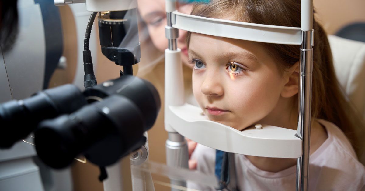
Visual Field Testing for Peripheral Vision

Read time: 6 minutes
We all know the profound impact that vision plays in our daily lives. While many of us take our central vision for granted, We often overlook our peripheral vision. One crucial aspect of regular eye examinations is visual field testing, a diagnostic tool that assesses the functionality and extent of a person's peripheral vision. In this comprehensive guide, we will delve into the historical context of visual field testing, explore contemporary insights, and discuss its significance in managing eye health.
Understanding Peripheral Vision
Peripheral vision, also known as side vision, refers to the ability to see objects and movement outside of the direct line of sight. It is the vision that occurs on the sides, above, and below the central field of view.
Your eyes have a wide field of vision, but only a small central area (called the fovea) provides sharp, clear vision used for tasks like reading or focusing on objects straight ahead. The peripheral vision surrounds this central vision and allows you to sense movement, light, shapes, and colors on the sides without directly looking at them.
Peripheral vision is facilitated by rod photoreceptor cells in the retina, which are more sensitive to motion and low light levels compared to the color-detecting cone cells concentrated in the fovea.
Good peripheral vision is important for activities like driving, playing sports, and maintaining full spatial awareness. It allows you to react to potential hazards or movement happening on the sides while your central vision is focused elsewhere.
However, the resolution and color perception decreases the further an object is from the center of gaze in the peripheral visual field. Things may appear blurred, distorted or desaturated in color toward the extreme sides of the visual field.
Many eye and neurological conditions can affect and limit peripheral vision. Visual field testing specifically evaluates the functionality and extent of the peripheral vision, helping detect issues early. Maintaining good peripheral awareness contributes to overall visual ability and safety.
Roots of Visual Field Testing
The concept of visual field testing can be traced back to the late 18th century when the Scottish ophthalmologist Thomas Reid pioneered the study of peripheral vision. Reid's work laid the foundation for further research and development in this area.
In the 19th century, significant advancements were made in the field of visual field testing. In 1856, a German ophthalmologist introduced the first systematic method for mapping the visual field, known as kinetic perimetry. This technique involved moving a small target from the periphery towards the center of the patient's vision, allowing for the mapping of the visual field boundaries.
As scientific knowledge and technological advancements progressed, more sophisticated methods of visual field testing emerged. In the late 19th century, static perimetry, a technique that involved presenting stationary targets at various locations within the visual field, was introduced.
The early 20th century witnessed the development of the first automated perimeters, which revolutionized visual field testing by providing more accurate and reproducible results. These early automated perimeters, such as the Goldmann perimeter and the Octopus perimeter, paved the way for the advanced technology we have today.
Current Advancements
In recent decades, visual field testing has undergone significant advancements, driven by technological innovations and a deeper understanding of various eye conditions. Contemporary insights have shed light on the importance of visual field testing in the early detection, diagnosis, and monitoring of several eye diseases, including glaucoma, neurological disorders, and retinal conditions.
Here are some key contemporary insights:
- Glaucoma is a progressive eye disease that can lead to permanent vision loss if left untreated. Visual field testing plays a crucial role in detecting and monitoring glaucoma-related vision loss, as it can identify defects in the peripheral vision caused by damage to the optic nerve.
- Another significant development is the integration of visual field testing into the management of neurological disorders, such as pituitary tumors, strokes, and multiple sclerosis. These conditions can affect the visual pathways and result in vision loss or visual field defects. Visual field testing can help diagnose and monitor these conditions, providing valuable information for treatment planning and follow-up care.
- Furthermore, visual field testing has become an essential tool in the evaluation and management of retinal conditions, such as age-related macular degeneration (AMD) and diabetic retinopathy. These conditions can cause central vision loss, and visual field testing can help determine the extent of peripheral vision remaining, guiding treatment decisions and monitoring progression.
Contemporary Visual Field Testing Techniques
Contemporary visual field testing techniques have evolved to provide more accurate, efficient, and user-friendly examinations. Some of the most commonly used techniques include:
- Standard Automated Perimetry (SAP): This is the most widely used visual field testing technique, particularly for glaucoma management. SAP utilizes an automated perimeter to present small light stimuli at various locations within the visual field. Patients are asked to press a button when they perceive the light stimulus, allowing for the mapping of their visual field.
- Frequency Doubling Technology (FDT): FDT is a specialized visual field testing technique that targets specific retinal ganglion cells, which are particularly susceptible to damage in glaucoma. This test can detect early glaucomatous changes and is often used in conjunction with SAP.
- Short Wavelength Automated Perimetry (SWAP): SWAP utilizes blue light stimuli to selectively test the function of specific retinal cells called short-wavelength-sensitive cones. This technique is particularly useful in detecting early glaucomatous changes and monitoring certain retinal conditions.
- Kinetic Perimetry: While less commonly used in routine clinical practice, kinetic perimetry remains a valuable tool in certain situations. It involves presenting a moving target from the periphery towards the center of the visual field, allowing for the mapping of visual field boundaries and the detection of specific defects.
- Electrophysiological Testing: In addition to traditional visual field testing techniques, electrophysiological tests, such as visual evoked potentials (VEPs) and electroretinography (ERG), can provide valuable information about the functional integrity of the visual pathways and retinal cells.
These contemporary techniques, combined with advanced imaging technologies like optical coherence tomography (OCT) and fundus photography, provide a comprehensive assessment of eye health and visual function.
The Importance of Visual Field Testing
Visual field testing plays a crucial role in the detection, diagnosis, and management of various eye conditions. Here are some key reasons why this examination is so important:
- Early Detection of Vision Loss: Visual field testing can detect subtle changes in peripheral vision that may be indicative of underlying eye conditions, such as glaucoma or neurological disorders. Early detection is essential for prompt treatment and preservation of vision.
- Monitoring Disease Progression: Regular visual field testing allows eye care professionals to monitor the progression of certain eye conditions over time. This information is valuable in determining the effectiveness of treatment and making necessary adjustments to the management plan.
- Comprehensive Eye Health Assessment: Visual field testing, combined with other diagnostic tests like OCT and fundus photography, provides a comprehensive assessment of eye health. This holistic approach helps identify and manage various eye conditions more effectively.
- Treatment Planning: The results of visual field testing can guide treatment decisions for conditions like glaucoma, neurological disorders, and retinal diseases. By understanding the extent and pattern of vision loss, your Urban Optiks Optometry eyecare professionals can tailor treatment plans to individual patient needs.
Visual field testing can help assess the impact of vision loss on a patient's daily activities and quality of life. This information is valuable in determining the need for supportive services, vision rehabilitation, or adaptive strategies.
The Takeaway
Visual field testing is a critical diagnostic tool in eye care, providing valuable insights into the functional integrity of the intricate visual system. Regular visual field testing is essential for early detection, disease monitoring, and personalized treatment planning. As we continue to push the boundaries of scientific knowledge and technological innovation, the future of visual field testing holds even greater promise.
Share this blog post on social or with a friend:
The information provided in this article is intended for general knowledge and educational purposes only and should not be construed as medical advice. It is strongly recommended to consult with an eye care professional for personalized recommendations and guidance regarding your individual needs and eye health concerns.
All of Urban Optiks Optometry's blog posts and articles contain information carefully curated from openly sourced materials available in the public domain. We strive to ensure the accuracy and relevance of the information provided. For a comprehensive understanding of our practices and to read our full disclosure statement, please click here.


